Home
/ Diagram Of Upper Chest Area / Sternum Wikipedia / The upper chest has two.
Diagram Of Upper Chest Area / Sternum Wikipedia / The upper chest has two.
Diagram Of Upper Chest Area / Sternum Wikipedia / The upper chest has two.. Check here to understand the function and part of it. Angina is the term for chest pain caused by poor blood flow to the heart. The chest wall thoracic lymph nodes receive drainage from the breasts, arms, pectoral muscles, and other muscles and skin located in the upper section of the chest. Powerful muscles that move the head and arms attach to these bones as well. There's only one acupuncture point on the chest.
Diagramme schnell und einfach erstellen. Chest pain has many possible causes, all of which need medical attention. So from one meathead to another let's go over the chest muscles themselves and what the chest is comprised of three separate muscles: Location of chest pain during angina or heart attack diagram. In other words, each area does something different.
Muscles Of The Chest And Upper Back from www.innerbody.com The epidermis is the outermost layer that provides a protective, waterproof seal over the body. the division into the separate, distinct parts of this muscle is about functionality. The bones of the chest — namely the rib cage and spine — protect vital organs from injury, and also provide structural support for the body. Understanding the basics of throat anatomy with diagram and pictures. It can feel like a heart attack. Profile view of female chest area. Rough area on the upper surface, where serratus anterior originates. See chest anatomy stock video clips.
Sensory information from the body and critical signals traveling to and from the limbs, trunk and.
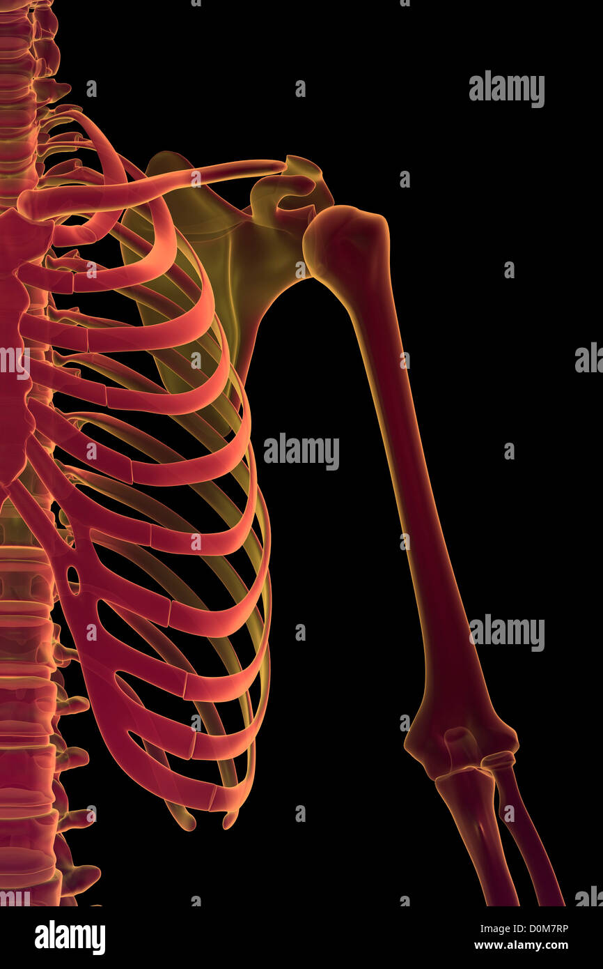
Location of chest pain during angina or heart attack diagram. The nervous system of the thorax is a vital part of the nervous system as a whole, as it includes the spinal cord, peripheral nerves, and autonomic ganglia that communicate with and control many vital organs. The upper chest has two. Nerves of the chest and upper back. Skeletal, cardiovascular, nervous and lymphatic systems. Anatomy of the chest and shoulder, anatomy of the chest organs, anatomy of the chest wall, anatomy of the chest wall and pleura, anatomy of upper chest area, human. I often get asked, how can i build thick powerful pecs? In the jaw area that draws. System respiratory respiratory organs of human body digestive and respiratory system medical chest internal structure of human body medicine body lungs biology intestines stomach anatomy torso human internal. The upper fibers, the middle fibers (called the middle trapezius), and the lower fibers (called the lower traps). Your pectoralis major and pectoralis minor muscles make up most of the muscle mass in your chest. In the diagram the square abcd is inscribed in circle o with diagonal ac 8. The sternum, commonly known as the breastbone, is a long, narrow flat bone that serves as the keystone of the rib cage and stabilizes the thoracic skeleton.
The upper chest has two. It forms the bulk of the chest area and can be easily seen on the surface in some people, for example weightlifters. It consists of the manubrium on its uppermost end, its body in the intermediate region and the small xiphoid process on its lowermost end. Anatomy of the chest and shoulder, anatomy of the chest organs, anatomy of the chest wall, anatomy of the chest wall and pleura, anatomy of upper chest area, human. the division into the separate, distinct parts of this muscle is about functionality.
Upper Chest High Resolution Stock Photography And Images Alamy from c8.alamy.com Doctors diagnose chest wall pain in at least 25% of patients who come to the emergency room for chest pain. Your pectoralis major and pectoralis minor muscles make up most of the muscle mass in your chest. Profile view of female chest area. As mentioned above, the trapezius muscle is divided into 3 areas: Nerves of the chest and upper back. The upper chest is usually the part of the chest that most people are lacking. Anatomy diagram rib area : It forms the bulk of the chest area and can be easily seen on the surface in some people, for example weightlifters.
These nodes drain the arm, the upper part of the chest wall comprising the breasts.
Illustration showing the lungs within the chest. Several muscles that move the arms, head, and neck have their origins on the sternum. Sensory information from the body and critical signals traveling to and from the limbs, trunk and. Skeletal, cardiovascular, nervous and lymphatic systems. Rough area on the upper surface, where serratus anterior originates. In this image, you will find an upper chest, substernal radiating to neck and jaw, substernal raiding down left arm, substernal radiating down left arm, epigastric radiating to neck, jaw, and arms, neck and jaw, left shoulder and down both arms, intrascapular in it. Chest wall pain is caused by problems affecting the muscles, bones and/or nerves of the chest wall. Chest muscles function in respiration while abdominal overall, these chest muscles start at the clavicle and insert at the sternum and the armpit area the chest muscle's main function is to bring the upper arm across the body. The sternum is a nearly flat rigid bone in the middle of human chest. There's two whole acupuncture points from the lung meridian located on the upper chest and collarbone area. The upper fibers, the middle fibers (called the middle trapezius), and the lower fibers (called the lower traps). Upper anterior muscles anatomy chest in computer tomography, color ct image. Skeletal, cardiovascular, nervous and lymphatic systems.
Webmd takes a look at the causes, symptoms, diagnosis, and treatment of this. The bones of the chest and upper back combine to form the strong, protective rib cage around the vital thoracic organs such as the heart and lungs. The circulatory system does most of its. The chest is the area of origin for many of the body's systems as it houses organs such as the heart, esophagus, trachea, lungs, and thoracic diaphragm. See chest anatomy stock video clips.
Muscles Of The Chest And Upper Back from www.innerbody.com Chest wall pain is caused by problems affecting the muscles, bones and/or nerves of the chest wall. Unfortunately, in many cases, that's as far as the doctor takes the diagnosis. Skeletal, cardiovascular, nervous and lymphatic systems. Location of chest pain during angina or heart attack diagram. A woman's chest — like the rest of her body — is covered with skin that has two layers. System respiratory respiratory organs of human body digestive and respiratory system medical chest internal structure of human body medicine body lungs biology intestines stomach anatomy torso human internal. Anatomy of the chest and shoulder, anatomy of the chest organs, anatomy of the chest wall, anatomy of the chest wall and pleura, anatomy of upper chest area, human. Man head and chest anatomy diagram with ghost effect.
Illustration showing the lungs within the chest.
In this image, you will find an upper chest, substernal radiating to neck and jaw, substernal raiding down left arm, substernal radiating down left arm, epigastric radiating to neck, jaw, and arms, neck and jaw, left shoulder and down both arms, intrascapular in it. Well now we have the answers for you, cep training revolutionary muscle growth secrets <== click here below is a diagram showing the chest muscles depicting where the different exercises target. There's two whole acupuncture points from the lung meridian located on the upper chest and collarbone area. Diagramme schnell und einfach erstellen. Lymph nodes of the lower limbs inguinal lymph nodes. The bones of the chest and upper back combine to form the strong, protective rib cage around the vital thoracic organs such as the heart and lungs. The major muscle in the chest is the pectoralis major. Nerves of the chest and upper back. As mentioned above, the trapezius muscle is divided into 3 areas: Webmd takes a look at the causes, symptoms, diagnosis, and treatment of this. The muscle has three heads giving it three points of. A woman's chest — like the rest of her body — is covered with skin that has two layers. The epidermis is the outermost layer that provides a protective, waterproof seal over the body.
